Powered By


 Continue with Facebook
Continue with Email
Continue with Facebook
Continue with Email
Ultrasound is an imaging test used to see real-time images of soft tissues and blood vessels. High-frequency sound waves build pictures so doctors can visualize what is happening inside the body. This technique is used alongside other tests to diagnose lymphoma.
There are two main types of lymphoma: Hodgkin lymphoma and non-Hodgkin lymphoma (NHL). Ultrasound is used primarily for diagnosing NHL where swollen lymph nodes are obvious, but it can be used as a tool in other diagnostic tests.
Ultrasound uses a special device known as a transducer, which sends sound waves into the body. These waves bounce off of organs and soft tissues. A computer program then turns the detected sound waves into pictures of the body. A radiologist analyzes the pictures to look for suspected abnormalities or to diagnose certain conditions.
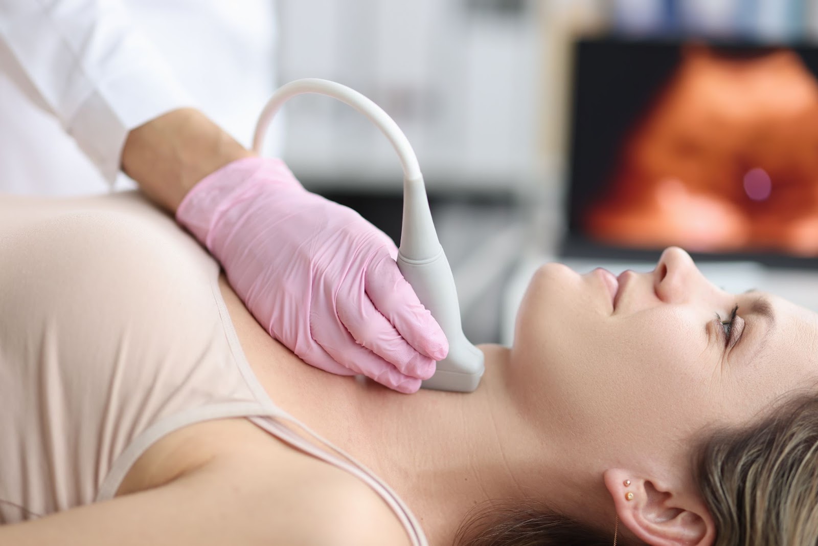
There are two types of ultrasound — external and internal. The most common type used for detecting lymphoma is the external ultrasound. It is used to take images of parts of the body that may be involved with lymphoma, including the neck, abdomen, or armpits. During an external ultrasound, a small amount of gel is placed onto the skin over the imaging area, and the transducer is placed onto this gel to help keep the images clear. The ultrasound technician (also known as a sonographer) may ask you to take a deep breath and hold still to get a better image during the scan.
Sometimes, an ultrasound requires the technician to apply pressure to the area for a better image. If this is uncomfortable, let them know. In total, the scan should take 15 to 30 minutes. Afterward, you can head home immediately.
Ultrasound is used to detect lymphoma and assist in diagnosing the disease. Ultrasound is just one of many imaging tests used for diagnosing lymphoma, including:
Along with a physical exam and biopsies, these tests help doctors confirm a lymphoma diagnosis and check what parts of your body are affected. This is the first step to receiving the right treatment for your lymphoma.
Lymphoma is a cancer of specialized immune cells known as lymphocytes. These white blood cells are found in the lymph nodes and other areas of the lymphatic system. Some lymph nodes, like the cervical lymph nodes in the neck, are close to the surface and can be easily felt and seen with ultrasound.
In suspected cases of lymphoma, ultrasound is used to visualize the lymph nodes in the neck. It can also detect enlarged lymph nodes in the abdomen that may be missed with other imaging techniques. These lymph nodes may press on other organs in the nearby area, such as the liver and kidneys.
Some people with NHL have swollen kidneys from urine blockage caused by enlarged lymph nodes. Lymphoma cells may also build up in the spleen, which causes it to swell and press on the stomach. Ultrasound can be used to evaluate the size and health of the kidneys and spleen in both Hodgkin lymphoma and NHL.
Biopsies are also used to diagnose lymphoma. This method takes a tissue sample from a swollen lymph node for lab testing. If the lymph nodes are close to the skin’s surface, your doctor can likely feel them enough to take a tissue sample correctly for the biopsy. However, in cases where the lymph nodes are deep in the body, such as the abdomen, they may use ultrasound to guide the biopsy. One study found this was an effective method for diagnosing both Hodgkin lymphoma and NHL.
Lumbar puncture is also used to diagnose lymphoma, in rare cases. This test takes a small amount of the fluid that surrounds the brain and spinal cord to look for lymphoma cells. Doctors can use ultrasound to guide them when they perform lumbar punctures. This helps reduce the number of attempts needed to place the needle.
In most cases, CT scans and PET scans are used to stage lymphoma (determine how far it has spread). Contrast dyes used in CT scans and radio-labeled glucose in PET scans contain very small amounts of radiation. While these levels are usually too low to cause any harm, many doctors avoid using the technique for pregnant people. If you are pregnant, be sure to discuss this with your doctor. Both ultrasound and MRI scans are generally safe during pregnancy.
Along with its usefulness as a diagnostic tool, ultrasound is also being studied for monitoring lymphoma in response to treatment. To see if a treatment is working or not, doctors will order PET/CT scans to see if the lymph nodes are shrinking. However, this method is not always ideal — some lymph nodes may shrink more slowly than others, which may make monitoring difficult. Treatment guidelines also recommend monitoring and restaging lymphoma after six to eight weeks of chemotherapy. In this amount of time, some lymphoma cases become resistant to the treatment and progress.
One small study looked at using different ultrasound techniques to monitor early treatment response from the first dose over the course of 30 days. The authors found that a specific type of ultrasound, known as B-mode ultrasound, provided a useful way to monitor how well participants with Hodgkin lymphoma or NHL responded to treatment. It helped doctors visualize the lymph nodes and measure their size throughout therapy to see which treatments were working and which were not.
Before an ultrasound, your doctor may give you some instructions. Depending on the area of the body being scanned, you may not be able to eat or drink for 12 hours before your scan. If the bladder and kidneys are being imaged, you may be asked to drink around 8 cups of water a few hours before the scan. This helps the bladder stand out from other organs in the images. If you are scheduled for an ultrasound, be sure to follow your doctor’s recommendations for the best way to prepare.
MyLymphomaTeam is the social network for people with lymphoma. More than 12,000 members come together to ask questions, give advice, and share their stories with others who understand life with lymphoma.
Have you had an ultrasound for your lymphoma? Share your experience in the comments below, or start a conversation by posting on MyLymphomaTeam.
Get updates directly to your inbox.


Can ultrasound be used to detect if PCMZBCL NHL, complete excision of 1 isolated tumour, 2022, no margins left, if cancer cells remain?
Continue with Facebook
Continue with Email
Continue with Facebook
Continue with Email
Become a member to get even more


 Join
Join


 Your Privacy Choices

This is a member-feature!
Sign up for free to view article comments.
We'd love to hear from you! Please share your name and email to post and read comments.
You'll also get the latest articles directly to your inbox.